Address
10 place de la Joliette
Atrium 10.4 – Etage 6
13002 Marseille, France
Name: Anthony SEBILLOT
University / Institution: Université de Rennes
Department / Research Group: Biosit (UMS/UAR CNRS 3480 – INSERM 018)
Position / Title: Research Engineer Inserm, Manager at H2P2 Core Facility (Histo
pathology High precision).
Research Area / Specialization: Histopathology
1. Please briefly describe your research focus and current projects.
H2P2 facility is a multidisciplinary core facility dedicated to advanced tissue analysis and high-resolution biological imaging. It provides a comprehensive environment for histological processing, histopathological evaluation, and phenotypic characterization of biological samples in both basic and translational research contexts. The platform integrates state-of-the-art instrumentation and expertise in tissue preparation, microscopy, and digital imaging, enabling high-quality morphological and molecular investigations.
The H2P2 facility performs histology work on animal and plant tissue. We bring our technical and technological skills in the various fields of histopathology.
We carry out tissue fixation, paraffin impregnation, thin sections with a microtome and cryostat, histology stainings (hematoxylin-eosin-saffron [HES], Sirius Red, Periodic acid-Schiff [PAS], Fontana-Masson, Oil red O staining, and many others stainings suitable for specific requests). An immunohistochemistry slide stainers (Ventana Discovery), designed specifically for research is available. This equipment can be used to improve the optimisation and reproducibility of immunohistochemical labelling, on a carrousel of 30 slides, each treated independently (choice of antigen unmasking buffer, temperature, and incubation time). This machine allows us to respond rapidly to the demands of users. This equipment has load rate of above 95%.
We also have extensive expertise in laser capture microdissection and we propose tissue studies by immunohistochemistry and fluorescent detection, in situ hybridisation, organotypic cultures (vibratome). We have expertise in immunolabelling (more than 1500 antibodies in our catalogue) as well as immunohistochemical staining: simplex ; multiplex staining, a technique that can highlight up to 6 different proteins per slide, using fluorescent or chromogenic markers ; hyperplex: actually more than 20 biomarkers to progress to 30 biomarkers in the next months).
We are developing Raman microspectroscopy imaging (Spontaneous/Stimulated Raman (SRS) on biological samples. We produce Tissue Microarrays (TMA) that can hold hundreds of tissues on a single microscope slide and we perform clarification (CUBIC/RapiClear) and marking of samples such as organoids, sections and entire organs.
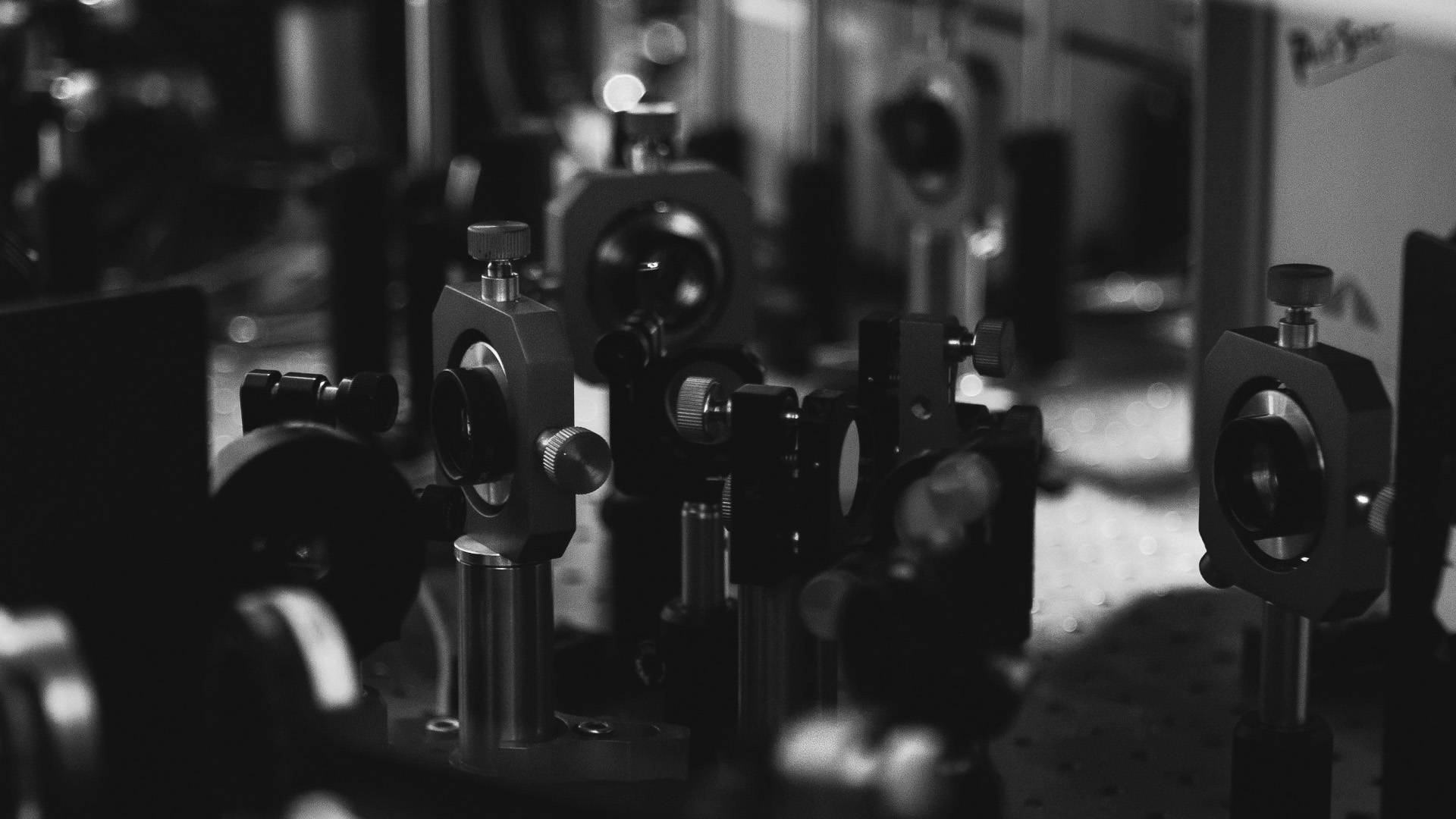
H2P2 also performed production virtual slides (brightfield and fluorescence imaging) and 2D image analysis (QuPath) and chemometrics. We visualise our slides using an epifluorescence microscope but also virtual slides from a Hamamatsu scanner with fluorescence. A confocal scanner, which is able to scan in depth, up to 300μm, with up to 7 different dyes. This scanner works also on brightfield mode. Its great interest is the ability to produce high resolution 3D images.
The equipment that equips the platform has been chosen to meet the needs of research laboratories. They offer great programming flexibility while providing reproducibility through automation.
2. What research needs or challenge were you aiming to address when looking for a
solution ?
We are constantly striving for improvement and progress. The H2P2 platform aim is to be able to continue to offer the cutting-edge technologies to research teams. Some devices/techniques are only available by the H2P2 team.
3. What factors influenced your decision to select our system ?
The choice of Lightcore Technologies’ BondXplorer for histopathological analysis was based on a combination of scientific and operational factors. The main criteria are likely the following:
Label-free tissue characterization
The BondXplorer enables molecular and structural imaging without the need for dyes or fluorescent markers. In histopathology, this feature is invaluable as it preserves tissue integrity, reduces preparation variability, and can accelerate the workflow compared to conventional staining methods.
High chemical specificity
H2P2 facility uses nonlinear optical and Raman imaging approaches that provide biochemical information directly from the tissue. This allows for the differentiation of tissue types, tumor margins, fibrosis, lipid distribution, and cellular architecture with molecular contrast superior to that of standard brightfield microscopy.
Reduced sample preparation time
Traditional histopathology often requires fixation, sectioning, staining, and multiple processing steps. A label-free optical workflow can shorten turnaround time and facilitate faster decision-making in research or translational medicine. Multimodal imaging capability: BondXplorer combines multiple imaging modalities within a single platform. For histopathological analysis, this enables the simultaneous acquisition of structural and molecular information, thus improving diagnostic reliability and tissue interpretation.
Multimodal imaging capability
BondXplorer combines multiple imaging modalities within a single platform. For histopathological analysis, this enables the simultaneous acquisition of structural and molecular information, thus improving the reliability of tissue analysis.
Preservation of native tissue state
Since chemical staining can alter biological samples, label-free imaging helps maintain the tissue closer to its native physiological condition. This is particularly important for research applications involving biomarker discovery.
Compatibility with digital pathology and AI workflows
Modern histopathology increasingly relies on quantitative image analysis and machine learning. The rich spectral and structural datasets generated by BondXplorer may support computational pathology and AI-assisted classification approaches.
Compact and user-friendly instrumentation
Lightcore emphasizes simplified deployment and reduced alignment complexity compared with traditional advanced optical systems. For a shared pathology or imaging facility, operational simplicity and lower maintenance requirements are major advantages.
Cost-efficiency over conventional high-end multimodal systems
The balance between imaging performance, versatility, size, and operating cost has made the BongXplorer attractive compared to larger or more specialized histopathology imaging platforms.
4. How has your experience been using our system in your research work ?
Our experience with the Lightcore Technologies BondXplorer system in our research has been extremely positive. This system has significantly improved the quality and efficiency of our histopathological analyses through label-free, high-resolution molecular imaging with remarkable ease of use. Its multimodal capabilities have allowed us to obtain rich structural and biochemical information from tissue samples, while reducing preparation time and preserving sample integrity. We were particularly impressed by the microscope’s ergonomics, which integrate seamlessly into our workflow and enable rapid acquisition of highly reproducible data. The advanced imaging performance of the BondXplorer has opened new avenues for more in-depth tissue characterization and has significantly enhanced the scientific value and impact of our research projects.
5. Have our system contributed to any improvements in your workflow, experiments,
efficiency, or research outcomes ?
The BondXplorer microscope has significantly improved our workflow, the efficiency of our experiments, and consequently, our research results. Its rapid, label-free imaging capabilities have significantly reduced sample preparation time and streamlined our histopathological analysis process, allowing us to analyze a larger number of samples with greater consistency and reliability. The high quality of the multimodal data generated by the system has improved the accuracy and depth of our tissue characterization, resulting in more robust findings. Furthermore, the platform’s intuitive interface and ease of use have facilitated training for our team and its daily operation. In summary, the BondXplorer has significantly increased our productivity, accelerated data acquisition and interpretation, and contributed to improved scientific quality across several ongoing research projects.
6. Are there any specific features or aspects you particularly value ?
We particularly value the exceptional label-free imaging capabilities of the BondXplorer microscope, which allow us to obtain highly detailed molecular and structural information while preserving the native state of tissue samples. The multimodal imaging approach is especially valuable, as it provides complementary biochemical and morphological insights within a single platform, greatly enhancing the depth of our analyses. We also highly appreciate the system’s ease of use, operational stability, and compact design, which make it well suited for routine use in a demanding research environment. In addition, the speed of image acquisition, the excellent reproducibility of results, and the overall quality of the generated data have been particularly impressive and have become major strengths in our research workflow.
The acquisition of the BondXplorer microscope represents a major strategic asset for the development of new collaborative projects and the strengthening of our innovation capabilities. This cutting-edge technology now enables us to respond much more competitively to national and European funding calls by proposing innovative approaches in histopathological imaging and label-free molecular analysis. The system also serves as a key driver for initiating and structuring multiple scientific and industrial collaborations with leading academic partners. Thanks to its advanced performance, versatility, and strong translational potential, the BondXplorer significantly enhances the attractiveness of our technological platform and opens new opportunities for multidisciplinary research projects with high scientific and applied impact.
7. Would you recommend our system to other researchers or institutions ? Why ?
Yes, I would highly recommend the BondXplorer system from Lightcore Technologies to other research institutions and laboratories. The system combines advanced label-free molecular imaging capabilities with excellent ease of use, and multimodal analysis performance, making it a highly valuable tool for histopathology and translational research. Its ability to generate high-quality structural and biochemical information while reducing sample preparation time has significantly improved our workflow efficiency, data reproducibility, and overall research productivity. In addition, the versatility and innovative nature of the microscope create strong opportunities for the development of collaborative projects, industrial partnerships, and competitive funding applications with leading academic institutions.
1 064
Finally, the Lightcore Technologies support team demonstrates exceptional expertise, responsiveness, and customer service, significantly enhancing the BondXplorer system user experience. Always available to answer technical and scientific questions, they ensure rapid problem resolution and minimize disruption to ongoing experiments. Beyond troubleshooting, they offer valuable guidance on optimizing system performance, adapting imaging protocols, and improving data quality, which directly impacts research results. Their proactive approach, strong scientific understanding, and commitment to user success make them a reliable and trusted partner for our research activities. In short, the quality and responsiveness of Lightcore’s support are major assets that greatly contribute to the seamless integration and effective use of the technology within our research platform.