Address
10 place de la Joliette
Atrium 10.4 – Etage 6
13002 Marseille, France
The BondXplorer™ microscope provides SRS imaging of both CH2 and CH3 bonds simultaneously. Using these images, we developed a post-processing virtual coloring method which imitates the H&E stained imaging commonly used in histopathology.
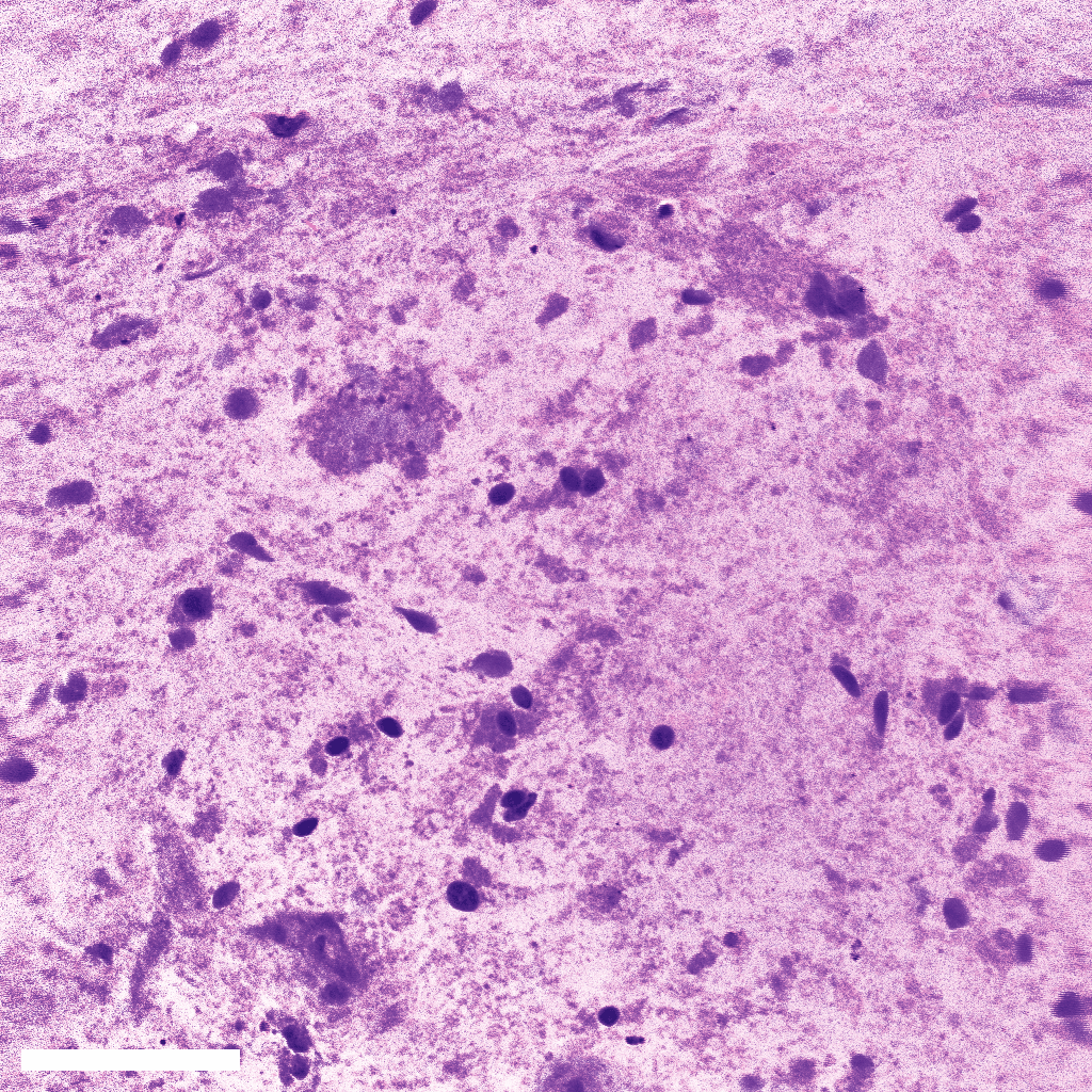
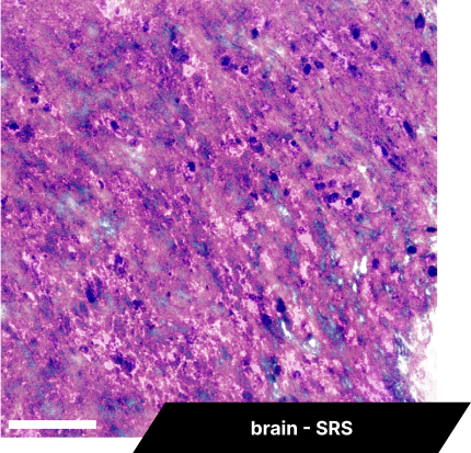
Brain SRS imaging
BondXplorer microscope
SRS imaging can reveal the internal structure of the brain, especially nucleus. Based on CH2 and CH3 bonds imaging, the BondXplorer™ microscope can produce an instantaneous label-free histological image, allowing to quickly distinguish a tumoral tissue from a healthy one. Z-stacks (see left image) and mosaics can be performed with this coloring, as well as live imaging. The scale bars are 85 and 50 µm for the left and right images respectively.
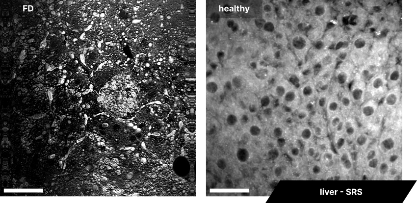
Human liver SRS imaging
BondXplorer microscope
The Bondxplorer™ microscope is designed to image SRS signal of both CH2 and CH3 bonds at the same time. CH3 bonds reveal areas with a lot of proteins, whereas CH2 bonds show mostly lipidic structures. Here we show CH2 bonds images of a healthy liver compared to a fat diet (FD) liver. The fat diet liver shows strong signal areas, which are lipid droplets, the hallmark of an unhealthy liver. The scale bars represent 43 µm.
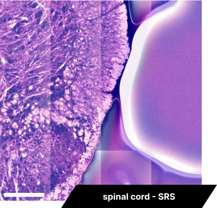
Spinal cord SRS imaging
BondXplorer microscope
Starting from SRS CH2 and CH3 bonds signal, we use our custom virtual coloring technique to produce a histology image of a spinal cord. In addition, the BondXplorer™ microscope can automatically scan a rather large area to produce a mosaic image, combination of multiple adjacent images acquired. Here is an example of a 6-by-6 colored mosaic image. The scale bar is 170 µm long.