Address
10 place de la Joliette
Atrium 10.4 – Etage 6
13002 Marseille, France
Our BondXplorer™ microscope and InSplorer™ endoscope offer a very wide range of imaging modalities with high resolution to address single cells in most biological tissues.
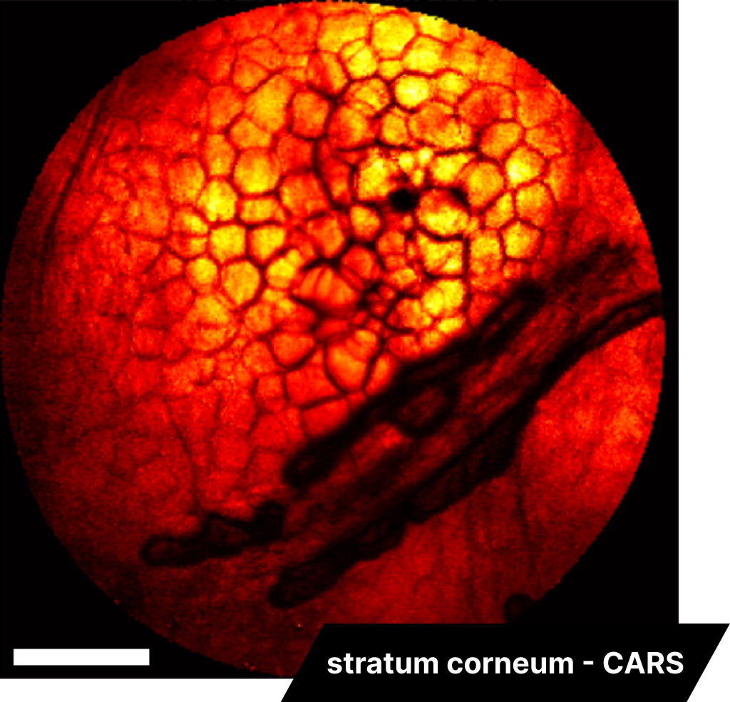
Stratum corneum CARS imaging
InSplorer endoscope
Coherent Anti-Stokes Raman Scattering (CARS) imaging is a powerful tool that enables the targeting of specific chemical bonds. It exhibits a particularly strong signal for CH bonds in lipids, around 2850 cm-1, making it highly beneficial for cellular imaging due to the lipid bilayer membrane of cells. An example of this can be seen in the imaging of a mouse skin sample (specifically, the stratum corneum layer) using the InSplorer™ endoscope. The scale bar in this context represents 120 µm.
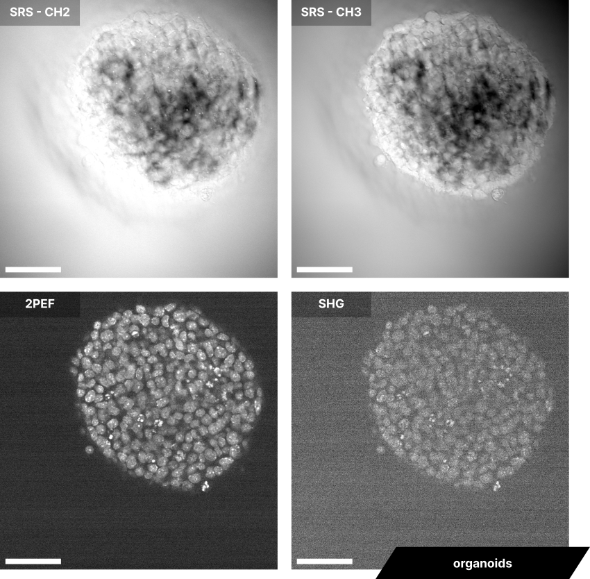
Organoids SRS, 2P and SHG imaging
BondXplorer microscope
An organoid is a cell-based in vitro model designed to replicate some functions of an organ. One of the main applications is for drug development. Our BondXplorer™ microscope system can perform imaging on an organoid with both SRS and multiphoton contrast. The present four images were captured simultaneously in an organoid sample using SRS of CH2-bond and CH3-bond, two-photon epifluorescence (2PEF) and Second Harmonic Generation (SHG). The organoid sample was prelabelled with fluorophore. The wavelengths utilized here are 792,5 nm for the pump, 1025 nm and 1034 nm for both Stokes beam. The scale bar is 50 µm.
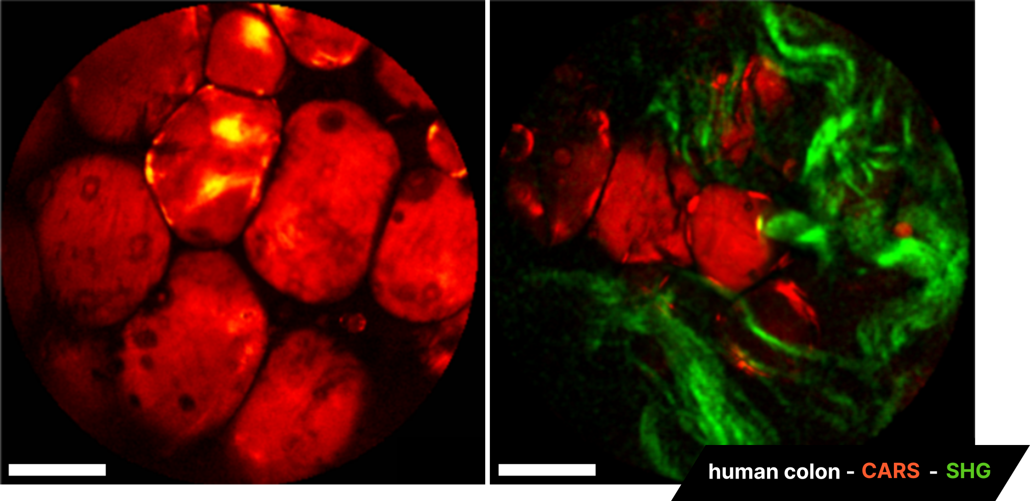
Colon CARS and SHG imaging
InSplorer endoscope
We conducted Coherent Anti-Stokes Raman Scattering (CARS) imaging on human colon biopsies to highlight the lipids present in the cell membrane. The multimodal capabilities of the endoscope also allow for the visualization of collagen fibers using Second Harmonic Generation (SHG) imaging. The scale bar represents 120 µm.
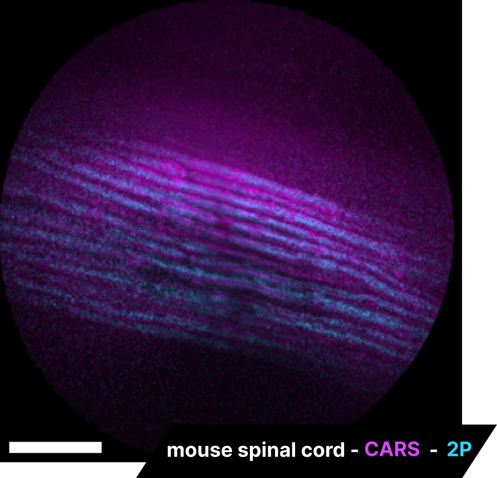
Spinal cord CARS and 2P imaging
InSplorer endoscope
Leveraging the multimodal capabilities of the InSplorer™ endoscope, we are able to image nervous system cells within a mouse spinal cord. Two-photon (2P) imaging enables us to visualize the spinal axons, while Coherent Anti-Stokes Raman Scattering (CARS) imaging accentuates the myelin membranes. The scale bar represents 120 µm.
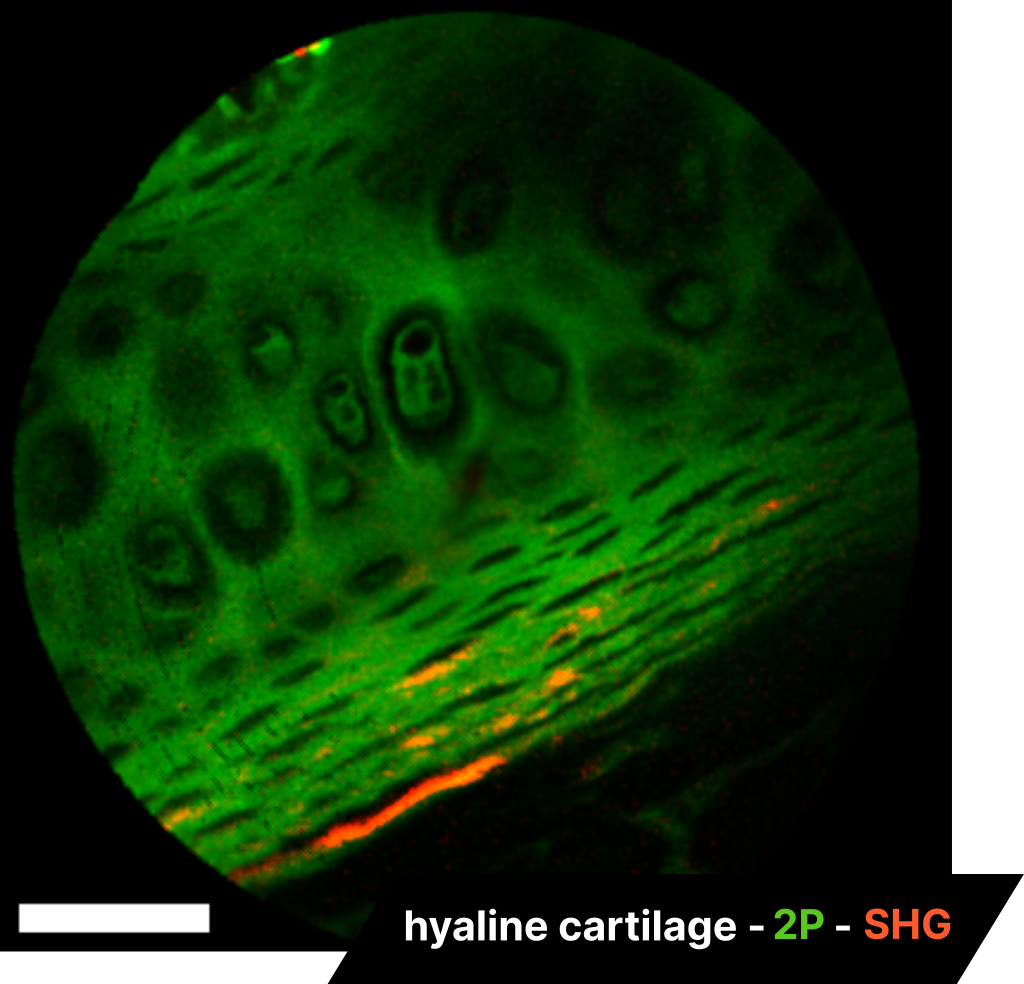
Cartilage 2P and SHG imaging
InSplorer endoscope
The image presented here provides an example of two-photon fluorescence imaging applied to cellular imaging, specifically in the case of a standard eosin-stained sample of hyaline cartilage. Due to the intrinsic nonlinear mechanisms of our endoscope, we are able to simultaneously provide Second Harmonic Generation (SHG) along with two-photon imaging. In this instance, it effectively highlights collagen fibers in addition to the cells. The scale bar is 60 µm.
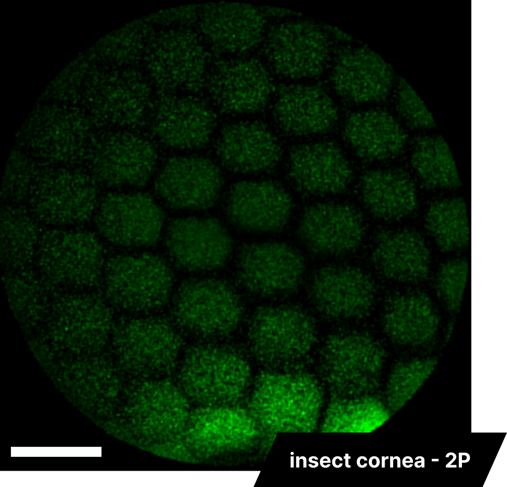
Insect cornea 2P imaging
InSplorer endoscope
The image on the right presents an endoscopic view of an insect cornea, showcasing the characteristic hexagonal shape of corneal endothelial cells. The scale bar in this image represents 60 µm.
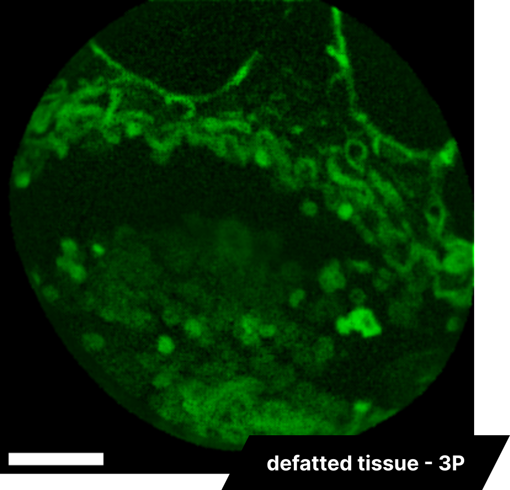
Defatted tissue 3P imaging
InSplorer endoscope
The InSplorer™ endoscope employs a fiber that delivers short laser pulses to a sample without temporal broadening. This feature allows for a high peak power on the samples, facilitating the use of three-photon (3P) imaging. The benefits of 3P imaging include higher resolution, optical sectioning, and increased penetration depth. An example of this can be seen in the application of 3P imaging to adipocyte imaging in defatted tissue. The scale bar in this image is 35 µm.