Address
10 place de la Joliette
Atrium 10.4 – Etage 6
13002 Marseille, France
Using our InSplorer™ endoscope and a custom hand-held endoscopic pen (Patent Pending), we are able to image the skin in real time, and up to a few hundreds of microns under the surface.
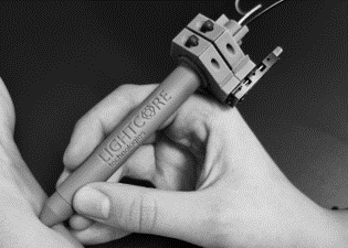
Custom pen endoscope
InSplorer endoscope
This innovative patented pen attachment transforms how you hold and maneuver the InSplorer™ endoscope, offering unparalleled control and comfort during imaging procedures. Equiped with a miniature translation stage, it allows to select the depth at which to image, as well as performing z-scan at the desired position.
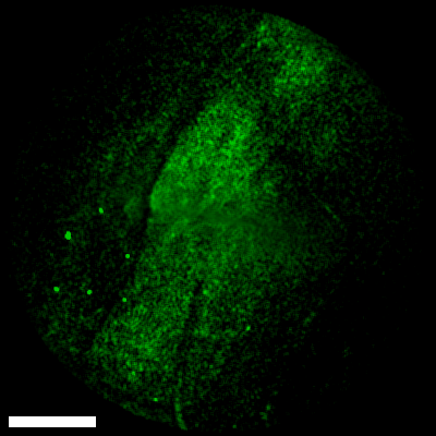
Surface mapping
InSplorer endoscope
Using our hand-held pen and with a high acquisition rate, we can easily move around the tissue to map out an area or look for a particular structure. In this example, we image the surface of the epidermis layer using 2-photon fluorescence at 6 FPS. We move along the arm for a total length of about 3 mm. The scale bar is 120 µm.
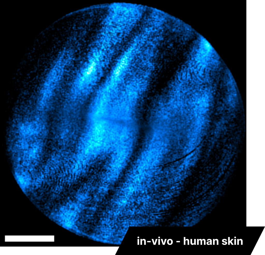
Collagen fibers imaging
InSplorer endoscope
The dermis layer of the skin can be reached to image collagen fibers using second harmonic generation (SHG). In this example, we image the human forearm, where the epidermis layer is thinner, to reach it, approximately 200-250 µm under the surface. It was acquired at 6 FPS, with excitation at 860 nm. The scale bar is 120 µm.
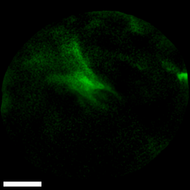
Z-scan
InSplorer endoscope
Similarly, using the same hand-held pen, we can hold it in place and scan the tissue in depth (along the z-axis). Here we image the surface of the epidermis layer using 2-photon fluorescence at 3 FPS and move the endoscope along the z-axis using the build-in stage of the pen. The scale bar is 60 µm.