Address
10 place de la Joliette
Atrium 10.4 – Etage 6
13002 Marseille, France
Nonlinear imaging, a technique that can be executed using our BondXplorer™ microscope or InSplorer™ endoscope, offers the ability to visualize a wide array of structures, including proteins present in biological tissues. A prime example is collagen, a well-known and abundant protein that is known to emit a robust second harmonic generation (SHG) signal. This advanced imaging approach allows us to explore and understand the intricate details of biological structures with remarkable precision.
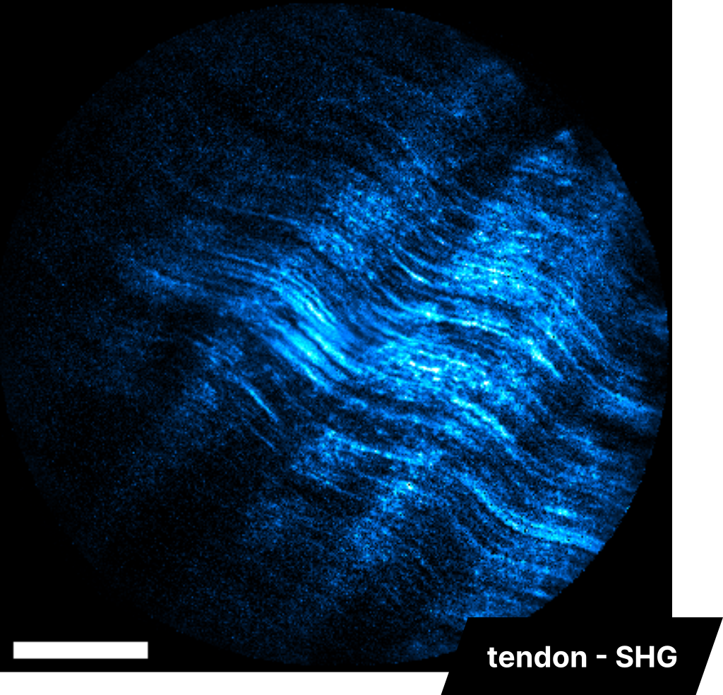
Tendon SHG imaging
InSplorer endoscope
The image on the left provides an illustrative example of endoscopic Second Harmonic Generation (SHG) imaging. It vividly displays the intricate network of collagen fibers present within a preserved tendon sample. The excitation wavelength employed for this imaging process is 920 nm. The scale bar in the image signifies a length of 60 µm, further emphasizing the high-resolution capabilities of our imaging technology.
Human bone SRS and SHG imaging
BondXplorer microscope
Bone is a collagen-abundant tissue with rich 3D structures. The BondXplorer™ microscope provides the z-stack function for all signal channels, meaning it automatically records several channels simultaneously then move along the z-axis to acquire several images according to the depth inside the tissue. Thus, the SRS imaging of CH3 bonds and SHG imaging combined with the z-stack function can be used to image the 3D structure of the bone, here a human rib cage bone. The scale bar in this example is 125 µm long.
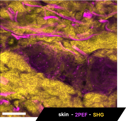
Skin SRS, 2P and SHG imaging
BondXplorer microscope
Apart from water, human skin is mostly made of protids and some of them can be revealed by fluorescence imaging. Here, we show an example of 2P and SHG imaging performed simultaneously on a human skin sample, using the BondXplorer™ microscope. 2P imaging reveals elastin whereas SHG reveals collagen, two of skin’s main proteins. The scale bar is 43 µm.
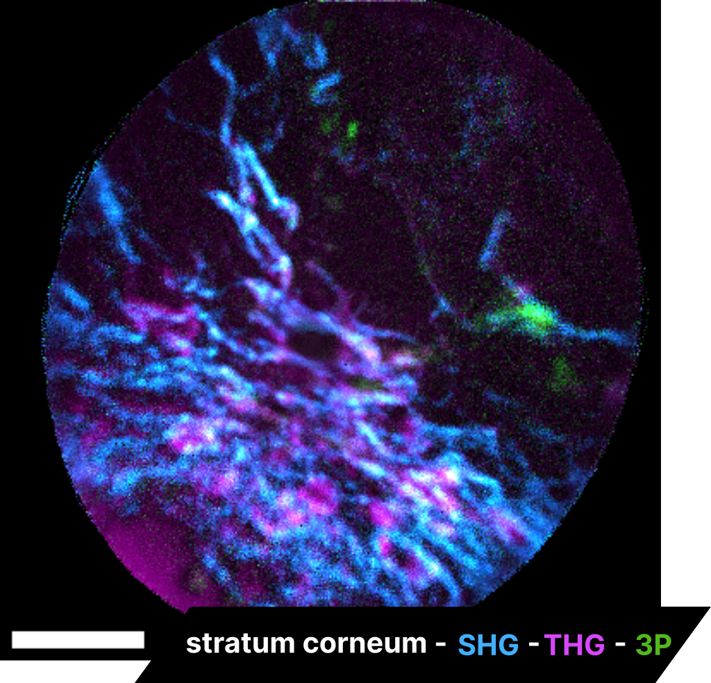
Skin SHG, THG and 3P imaging
InSplorer endoscope
We are able to integrate Second Harmonic Generation (SHG) imaging of collagen fibers with cellular imaging using a distinct imaging modality, highlighting the multimodal capabilities of the InSplorer™ endoscope. In this particular instance, the procedure is performed on a mouse skin sample, with cellular imaging achieved through Third Harmonic Generation (THG) and 3-photon (3P) autofluorescence. The scale bar represents 50 µm.
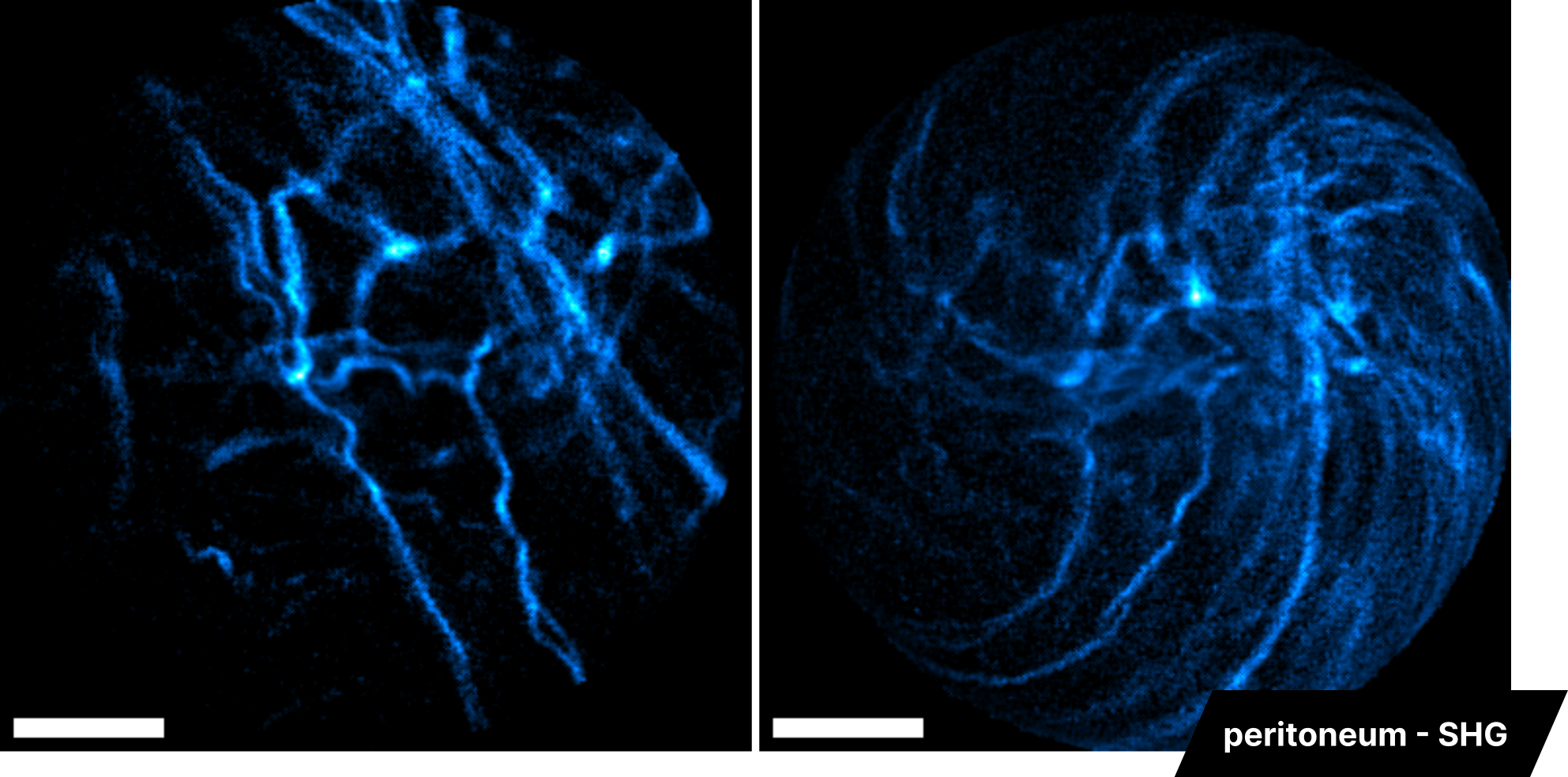
Peritoneum SHG imaging
InSplorer endoscope
In a similar vein, we conducted Second Harmonic Generation (SHG) imaging on a preserved peritoneum sample using the InSplorer™ endoscope. This process effectively illuminates the structure of collagen fibers within the tissue. The excitation wavelength used is 860 nm, and the scale bars represent 60 and 70 µm respectively.