Address
10 place de la Joliette
Atrium 10.4 – Etage 6
13002 Marseille, France
Investigating neuronal activity necessitates real-time brain imaging with a high acquisition rate. Our InSplorer™ endoscope, with its compact size and lightweight design, is capable of accessing the brain for in-vivo interrogation at speeds of up to 10 FPS. This makes it particularly suitable for monitoring calcium activity in neuronal cells in-vivo.
Real-time neurons activity
InSplorer endoscope
The videos on the left showcase real-time two-photon imaging of mice brains that have been genetically altered to enhance neuronal responses. Here, we present examples of real-time imaging performed at 6 and 8 FPS respectively. The calcium imaged exhibits a bright response to external stimuli, effectively highlighting neurons and dendrites. The scale bars represent 60 and 50 µm respectively. These experiments were conducted at the Institut de Neurologie de la Méditerranée (Inmed) in Marseille, France, in accordance with EU ethical rules on animal research.
Freely moving mouse brain imaging
We have designed a custom helmet that attaches the InSplorer™ distal head to the mouse skull. The helmet itself weighs 1 gram, and the overall weight on the head is just about 3.5 grams. This design allows the mouse to move freely with the endoscope attached, enabling us to image the brain in real-time. This setup fully leverages the capabilities of in-vivo endoscopic imaging.
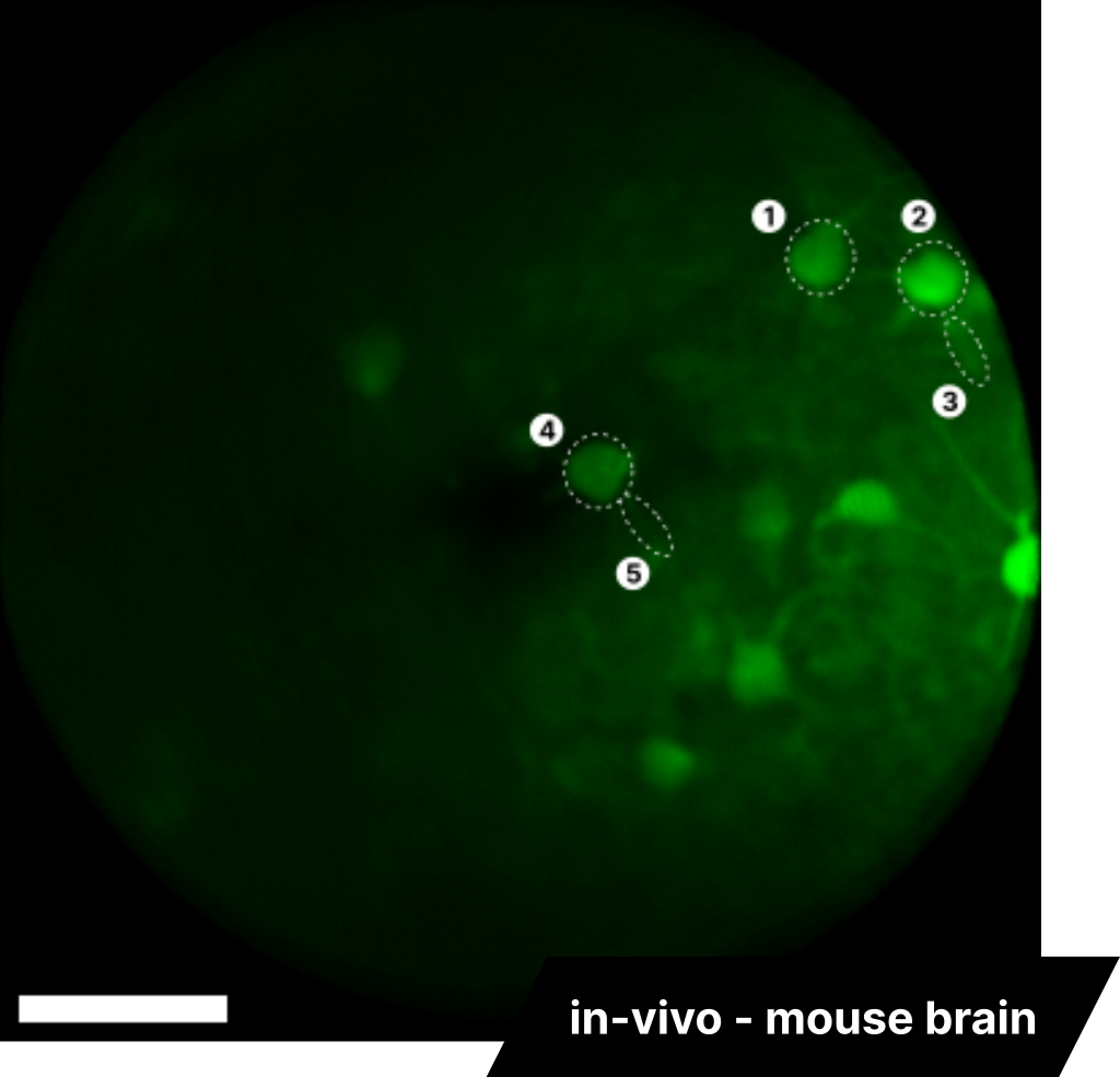
Neurons and dendrites activity tracking
InSplorer endoscope
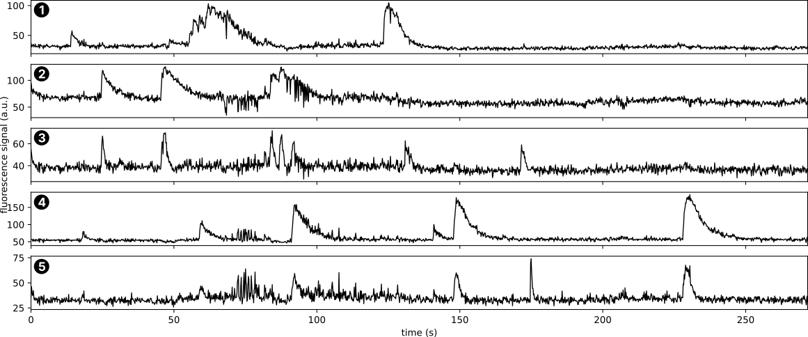
By harnessing the capabilities of the endoscope, we can monitor the activity of individual cells or even dendrites. The image here is a projection of all images captured during an imaging session, showcasing all cells and dendrites that exhibited activity. By selecting specific cells and dendrites, we can extract their activity patterns and correlate them with the mouse’s movements and/or interactions. This is illustrated in the example graphs on the right.